Have you ever wondered how doctors plan precise cancer treatments? CT simulation plays a critical role in radiation therapy by creating detailed 3D images of the body. This advanced technology ensures that radiation is delivered accurately to target tumors, minimizing damage to surrounding healthy tissue.
In this post, we’ll explore what CT simulation is, its importance in radiation therapy, and how it improves the accuracy of cancer treatment. You'll learn about the role of a computed tomography simulator and how CT simulators in radiotherapy are transforming cancer care.
CT simulation is a specialized imaging technique used to create a 3D model of a patient’s body. Unlike traditional CT scans, which are typically used for diagnostic purposes, CT simulation is specifically designed for treatment planning in radiation therapy. The process involves detailed imaging that helps clinicians visualize tumors and surrounding tissues, ensuring precise targeting for radiation treatment.
The main goal of CT simulation is to help plan the most accurate and effective radiation therapy. With the use of a computed tomography simulator, doctors can define the exact location, shape, and size of the tumor. This enables precise radiation delivery while minimizing exposure to healthy tissue. CT simulation is essential in radiotherapy as it provides the foundation for tailoring treatment plans for each patient.
CT Simulator in Radiotherapy: By using a CT simulator, doctors can improve the effectiveness of radiation treatment, ensuring that it reaches the tumor while protecting healthy organs.
A CT simulation involves several key steps to ensure accurate radiation therapy planning. Here's a quick breakdown:
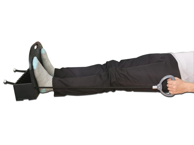
Patient Positioning: The first step is positioning the patient on the CT simulator table. Using custom-made immobilization devices, the patient remains still throughout the procedure. This ensures the tumor is consistently targeted during radiation treatment.
Imaging Process: During the CT simulation process, the patient undergoes a scan, where multiple X-ray images are taken. These images are then combined to create a 3D map of the body. This map shows the tumor’s exact location, size, and shape. The data gathered helps the doctor plan where to focus radiation.
The CT simulation works by providing detailed images that guide doctors in planning radiation therapy. Using the CT simulator, they ensure the radiation targets the tumor precisely, minimizing damage to healthy tissue.
CT simulation is a cornerstone of modern radiation therapy. It helps plan the precise delivery of radiation to cancerous tumors while minimizing damage to healthy surrounding tissue. By using a computed tomography simulator, radiation oncologists can create detailed 3D images that guide them in designing an accurate treatment plan.
One of the key reasons CT simulation is crucial for radiation therapy is its image accuracy. The high-resolution 3D images created during the CT simulation process allow doctors to pinpoint the tumor's exact size, shape, and location. This ensures radiation is focused on the tumor, significantly improving treatment effectiveness and reducing the risk of side effects to healthy tissue.
The CT simulation for radiation therapy allows for a customized approach to treatment, optimizing the chances of success while minimizing unnecessary exposure.

The primary piece of equipment used in CT simulation is the CT simulator itself, a specialized machine designed to capture detailed 3D images of the patient’s body. The CT scanner within the simulator takes multiple X-ray images, which are then processed to create a 3D map of the body. This map helps oncologists plan radiation treatment accurately.
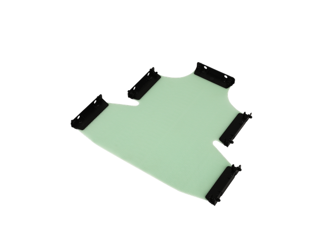
Additionally, laser positioning systems are used to align the patient properly, ensuring that the tumor is targeted precisely during radiation therapy. To prevent any movement during the procedure, immobilization devices are used to hold the patient in the same position throughout the simulation.
Modern CT simulators come equipped with advanced features, such as 4D CT simulation. This technology allows clinicians to account for tumor movement, especially in areas like the lungs, where motion due to breathing is a factor. 4D CT simulation improves the accuracy of radiation treatment by tracking tumor changes in real-time.
The CT simulation equipment continually evolves, incorporating these new technologies to enhance precision and treatment effectiveness.
CT simulation plays a crucial role in tumor localization by providing highly detailed 3D images of the patient's body. The CT scanner captures multiple X-ray images from different angles, which are then combined to create a 3D model. This model allows oncologists to pinpoint the exact location, size, and shape of the tumor. The CT simulator in radiotherapy ensures that radiation treatment targets the tumor precisely, minimizing harm to surrounding healthy tissue.
The primary clinical benefit of tumor identification with CT scan is the increased accuracy in radiation therapy. With CT simulation for radiation, the precise positioning of radiation beams is achieved. This leads to improved treatment outcomes, as the radiation is delivered exactly where it is needed, reducing the risk of side effects. The ability to accurately identify and locate tumors ensures that radiation is most effective and has the least impact on healthy organs and tissues.
While both a CT scan and a CT simulation use similar technology, their purposes and applications differ significantly.
CT Scan: A regular CT scan is primarily used for diagnostic purposes. It creates detailed images of organs and tissues to help doctors identify diseases, injuries, or abnormalities. The focus is on providing quick and accurate images for diagnosis.
CT Simulation: A CT simulation is a specialized procedure used in radiation therapy. It helps create a 3D map of the body, allowing doctors to plan radiation treatment precisely. The main goal is not diagnosis but to accurately target tumors and protect healthy tissue.
Purpose: A CT scan is for diagnosis, while CT simulation is for treatment planning.
Application: CT scans are used in routine medical checkups, while CT simulations are specific to radiation therapy.
Technology: Both use similar technology, but CT simulations include additional features, such as 3D imaging and immobilization devices, which aren’t typically needed in regular CT scans.
By comparing CT scan vs CT simulation, it’s clear that while both use imaging, their goals and methods differ based on the intended outcome.
CT simulation is essential for patients undergoing cancer treatment, especially those who require radiation therapy. It is commonly used for various types of cancer, such as:
Lung Cancer: Tumors in the lungs need precise targeting due to nearby critical organs.
Prostate Cancer: Accurate mapping of the prostate area helps in effective radiation therapy.
Head and Neck Cancers: Tumors in these regions require careful planning to avoid damage to delicate structures.
Other conditions, like brain tumors or certain types of soft tissue cancers, also benefit from CT simulation for cancer treatment.
CT simulation is primarily used when radiation therapy is needed. It ensures that radiation is precisely directed at the tumor. This process is vital for patients with tumors in difficult-to-reach areas or those near critical organs. CT simulators are often used before starting the radiation treatment to plan the safest and most effective approach.
In short, CT simulation is a key tool in radiation therapy, ensuring accurate tumor targeting for various types of cancer.
Once the CT simulation is complete, the data is used to create a precise radiation therapy plan. The process begins with the oncologist reviewing the 3D images generated by the computed tomography simulator. These images provide a detailed view of the tumor's location, size, and surrounding tissues. Using this information, the treatment team can outline the tumor and set boundaries for radiation. This ensures that radiation is directed accurately to the tumor while avoiding healthy tissues.
The oncologist, medical physicist, and dosimetrist work together to design a treatment plan based on the CT simulation data, ensuring optimal results.
The precise data from CT simulation for radiation helps optimize the delivery of radiation, minimizing side effects. By pinpointing the exact location of the tumor, doctors can carefully plan the radiation beams' path. This reduces the risk of damaging surrounding healthy tissue, ensuring more effective treatment. CT simulators in radiotherapy also enable personalized treatment, adapting the radiation therapy plan based on the patient’s specific anatomy and tumor characteristics.
With CT simulation for treatment planning, doctors can significantly improve the accuracy and safety of radiation therapy, making it more effective for patients.
One of the main benefits of CT simulation is its ability to improve the accuracy of radiation therapy. By creating detailed 3D images of the body, the CT simulator allows doctors to precisely locate the tumor and surrounding tissues. This helps ensure radiation is delivered directly to the tumor, minimizing the risk of exposure to healthy tissue. CT simulation for radiation is crucial for fine-tuning treatment plans, making them more effective.
CT simulation significantly enhances patient safety during radiation therapy. By accurately mapping the tumor and vital organs, the radiation therapy team can precisely target the tumor, reducing the risk of damaging nearby healthy tissues. The detailed images from the computed tomography simulator enable better protection for sensitive areas, such as the heart or lungs, ensuring that radiation only affects the intended area.
Comfort during the CT simulation process is another key advantage. Immobilization devices are used to keep the patient still during the simulation, ensuring accurate positioning for radiation treatment. These devices help patients remain in the same position throughout the process, increasing both comfort and treatment accuracy. CT simulation in radiotherapy is designed to minimize discomfort while ensuring that the treatment is precise and effective.
By improving accuracy, safety, and comfort, the advantages of CT simulation make it a crucial part of the radiation therapy process.
One concern related to CT simulation is the exposure to radiation. While the amount of radiation used in a CT simulator is typically low, it’s still a factor to consider. The patient is exposed to a small dose of radiation during the simulation process, similar to the radiation levels in a traditional CT scan. However, the benefits of using CT simulation for radiation treatment planning generally outweigh this risk, as the radiation exposure is essential for accurate tumor localization.
Although CT simulation for radiation offers many advantages, it has some limitations. For example, it may not be able to detect certain types of tumors, particularly very small or deeply located ones, that might require additional imaging techniques. CT simulators are also limited when it comes to visualizing soft tissues in detail. While CT scans are great for viewing bones and organs, other imaging methods like MRI may be used in conjunction to get a clearer picture of soft tissue structures.
Despite these CT simulation limitations, the technology remains an essential part of planning radiation therapy. It provides a high level of accuracy, though it may need to be complemented with other diagnostic tools for a comprehensive treatment plan.
A typical CT simulation session usually takes about 30 to 45 minutes. The exact duration may vary depending on the complexity of the treatment plan and the need for any adjustments during the process. While the actual imaging only takes a few minutes, positioning the patient and ensuring everything is aligned properly can take additional time.
During the CT simulation for radiation, patients can expect to lie still on a treatment table. Immobilization devices may be used to keep the body in the exact position for precise imaging. Patients will not feel any pain, but they may need to stay still for the duration of the procedure. The CT scanner will rotate around them, capturing images to create a 3D model for treatment planning.
Overall, CT simulation duration is generally short, and the process is designed to ensure accuracy and comfort for the patient.
One of the great advantages of CT simulation is that it is a completely non-invasive and painless procedure. Unlike other medical procedures, CT simulation for radiation does not involve any needles, incisions, or uncomfortable interventions. Patients simply lie still while the computed tomography simulator takes a series of detailed images. There is no pain involved during the scan itself.
During the CT simulation, patient comfort is a top priority. Immobilization devices are used to help patients stay in the same position, which ensures accurate results while minimizing discomfort. These devices are specially designed to be as comfortable as possible, and patients are instructed to relax during the process. The entire procedure is relatively quick, and many patients report feeling at ease throughout the session.
So, is CT simulation painful? No, it is a pain-free process aimed at ensuring both comfort and accuracy during treatment planning.
One of the most important advantages of CT simulation is its accuracy in targeting tumors for radiation treatment. The CT simulator provides detailed, high-resolution 3D images of the body, allowing oncologists to pinpoint the exact location, size, and shape of a tumor. This precision ensures that radiation is directed exactly where it's needed, reducing the risk of irradiating healthy tissue.
Clinical studies consistently show the effectiveness of CT simulation for radiation in improving treatment accuracy. The accuracy of CT simulation is critical in planning radiation therapy, especially for tumors located near sensitive organs. Studies have shown that using CT simulation for treatment planning leads to better outcomes by minimizing damage to surrounding tissues. Additionally, the detailed images allow doctors to customize treatment plans based on the patient's specific anatomy.
The CT simulation for precise treatment has proven to be a game-changer in ensuring targeted, effective radiation therapy with minimal side effects.
4D CT simulation is an advanced technique used in radiation therapy to track tumor movement, particularly during breathing. Unlike standard CT simulation, which captures static images, 4D CT simulation takes multiple images over time, creating a dynamic, moving 3D model. This allows oncologists to account for changes in tumor position caused by the patient’s natural respiration, especially in areas like the lungs, where tumors can shift with each breath.
The primary advantage of 4D CT in radiation therapy is its ability to improve the precision of radiation treatment for tumors that move, such as those in the lungs or abdomen. With 4D CT simulation, radiation therapy plans can be adjusted to target tumors throughout their movement cycle, ensuring accurate delivery of radiation even when the tumor is not stationary. This reduces the risk of radiation exposure to surrounding healthy tissue and improves the effectiveness of the treatment.
In short, 4D CT simulation provides a more comprehensive and dynamic approach to radiation therapy, offering better outcomes for tumors affected by respiration.
After a CT simulation, the collected data is used to create a personalized treatment plan for radiation therapy. The images and 3D model generated during the CT simulator session are analyzed by the oncologist, dosimetrist, and medical physicist. Using this data, they will plan the precise location, angle, and dosage of radiation needed to target the tumor effectively. This ensures that the tumor receives the maximum dose while protecting surrounding healthy tissues.
Once the radiation therapy plan is developed, the next steps involve reviewing and finalizing the treatment. The patient may undergo further consultations or imaging to confirm the plan's accuracy. In some cases, the treatment plan is adjusted based on new information or any changes in the tumor’s size or location. After CT simulation, the patient typically begins radiation treatment, following the carefully planned schedule.
After CT simulation, the process moves from planning to actual treatment, ensuring every step is tailored to deliver the best possible outcome.
CT simulation plays a vital role in radiation therapy by improving the accuracy of cancer treatments. It provides detailed 3D imaging, allowing oncologists to pinpoint tumors and plan radiation delivery precisely. This results in more effective treatment and minimizes harm to healthy tissues.
By using a computed tomography simulator, CT simulation for radiation ensures safer, more accurate treatments. It is an essential tool in modern cancer care, enhancing the precision and effectiveness of radiation therapy, and ultimately improving patient outcomes.
A: No, CT simulation is not the same as a PET scan. While both use imaging technology, CT simulation is specifically used for planning radiation therapy, creating detailed 3D maps of the body. PET scans are used for detecting metabolic activity and identifying cancer cells.
A: CT simulation is beneficial for many types of cancer, especially those requiring radiation therapy, such as lung, prostate, and head and neck cancers. However, it may be complemented by other imaging methods for certain tumors.
A: CT simulation is typically performed once before starting radiation therapy to plan the treatment. If the tumor changes in size or location, a follow-up simulation may be required.
 English
 简体中文
简体中文