Views: 0 Author: Site Editor Publish Time: 2025-08-13 Origin: Site

CT Simulation positions and immobilizes you for accurate tumor targeting.
It allows doctors to map tumors and healthy tissues in multiple views.
Digital data from ct simulation improves treatment planning and safety.
Advanced ct techniques enhance tumor definition, leading to better outcomes.
A CT scan helps doctors diagnose medical conditions by creating detailed images of your body quickly.
CT simulation prepares you for radiation therapy by mapping your body in the exact position for treatment.
CT simulation uses special devices to keep you still and marks your skin to guide precise radiation targeting.
You prepare differently for each: CT scans require removing metal and may use contrast dye, while CT simulation needs careful positioning and sometimes bladder or bowel preparation.
CT simulation machines have larger openings and flat tables to fit immobilization devices and match treatment setups.
CT scans are fast and used for many conditions like injuries, cancers, and organ problems, while CT simulation focuses on planning safe and accurate cancer treatment.
Your care team uses CT simulation images to design personalized radiation plans that protect healthy tissue and target tumors.
Asking your care team questions about preparation, procedure, and results helps you feel confident and involved in your care.

You may undergo a ct scan for several important reasons. Doctors use ct imaging to diagnose and evaluate a wide range of medical conditions.
You might need a ct scan to detect bone fractures or deformities.
Physicians rely on ct scans to identify cancers and locate tumors.
A ct scan helps assess heart conditions, kidney irregularities, and kidney stones.
You may need this test to diagnose lung diseases, pulmonary embolism, or osteoporosis.
After an accident, a ct scan can reveal internal injuries or bleeding.
Doctors use ct scans to guide procedures such as biopsies and monitor treatment effectiveness.
Tip: If your doctor orders a ct scan, ask about the specific reason and what information they hope to gain.
A ct scan uses advanced X-ray technology and computer algorithms to create detailed images of your body.
The ct scanner emits X-rays from multiple angles around you.
Detectors measure how much X-ray passes through your tissues.
Dense tissues like bone absorb more X-rays, while less dense tissues like lungs absorb less.
The system assigns a Hounsfield Unit to each tissue based on its density.
Computer algorithms reconstruct these measurements into cross-sectional images.
Modern ct scanners use spiral and multislice techniques for faster scans and higher resolution.
Some ct scans use contrast agents to highlight specific structures or blood vessels.
Note: Spectral ct technology can differentiate tissue types more precisely by using energy-dependent information.
You prepare for a ct scan by following several steps.
Remove all metallic objects such as jewelry, watches, and hearing aids.
Change into a hospital gown if requested.
If your scan requires contrast dye, a technician inserts an IV catheter.
Review any allergies or special instructions with your care team.
You lie on a long, narrow table that slides into the circular ct scanner.
The technician may secure you with straps to keep you still.
The technician leaves the room and operates the scanner remotely, communicating with you through an intercom.
The scanner rotates around you, producing noise as it captures images.
You may need to hold your breath or remain very still to avoid blurry images.
The scan usually takes between 20 minutes and 1 hour.
Once the ct scan finishes, you can resume normal activities unless you received contrast dye.
The images go to a radiologist for interpretation.
Your doctor reviews the results and discusses them with you.
If you had an IV, the technician removes it and checks for any reactions.
Tip: Ask your doctor when you will receive your ct scan results and what the next steps will be.
You will find that ct scans play a vital role in many areas of medicine. Doctors rely on ct imaging to quickly and accurately diagnose a wide range of conditions. Here are some of the most common uses:
Trauma and Injury Assessment: You may need a ct scan after an accident to check for fractures, internal bleeding, or head injuries.
Neurological Imaging: Doctors use ct scans to detect strokes, brain tumors, aneurysms, and other brain injuries.
Cardiovascular Evaluation: A ct scan can help your doctor assess coronary artery disease, aneurysms, and vascular malformations.
Pulmonary Imaging: If you have symptoms of lung cancer, pulmonary embolism, or pneumonia, a ct scan provides detailed images of your lungs.
Abdominal and Pelvic Examination: Ct scans help identify tumors, cysts, inflammation, and gastrointestinal disorders in your abdomen and pelvis.
Oncological Staging and Monitoring: Your care team may use ct scans to determine the size, location, and spread of tumors, which is essential for cancer treatment planning.
Musculoskeletal Imaging: Ct scans reveal fractures, arthritis, and soft tissue injuries that may not show up on regular X-rays.
Vascular Imaging: Doctors use ct angiography to detect aneurysms, dissections, and blocked arteries.
Infection Detection: Ct scans can locate abscesses or collections of pus in your body.
Procedural Guidance: Your doctor may use ct imaging to guide biopsies, drainages, or surgical planning.
Screening: Ct scans screen for diseases such as lung cancer in high-risk individuals or abdominal aortic aneurysms.
Note: Ct scans support specialists in orthopedics, neurology, cardiology, oncology, pulmonology, and general surgery. This versatility makes ct a cornerstone of modern medical imaging.
You benefit from ct scan technology because it combines speed, detail, and versatility. The following table highlights how ct scans compare to other imaging methods:
Imaging Modality | CT Scan Features | Comparison Highlights |
|---|---|---|
CT Scan | Uses X-rays and computer processing to create detailed 3D cross-sectional images; rapid acquisition; excellent spatial resolution; good bone imaging; involves ionizing radiation | Provides structural imaging with fast scan times; excels in skeletal, thoracic, and oncological imaging; lower soft tissue contrast than MRI; higher radiation than X-ray and ultrasound |
MRI | Uses magnetic fields and radio waves; excellent soft tissue contrast; no ionizing radiation | Slower scan times than ct; better soft tissue detail; no radiation exposure |
X-ray | Produces 2D images using X-rays; quick and widely available | Less detailed than ct; 2D only; lower radiation dose than ct |
Ultrasound | Uses sound waves; real-time imaging; no radiation | Lower resolution; limited penetration; best for soft tissues and fluid-filled structures |
PET/SPECT | Uses radioactive tracers to image metabolic and functional activity | Functional/metabolic imaging rather than structural; combined with ct for anatomical reference |
Ct scans stand out for their ability to produce fast, detailed 3D images of your body. You can expect excellent visualization of bones, lungs, and tumors. While ct uses more radiation than standard X-rays, it provides much more information in a single scan. Doctors often choose ct when they need rapid answers or detailed structural images.

You undergo ct simulation when your care team needs to plan precise radiation therapy. The main goal is to create a map of your internal anatomy in the exact position you will hold during treatment. Ct simulation helps your doctors contour tumors and healthy organs, so they can target cancer while protecting normal tissue. This process uses a ct simulator to produce 3-D images that guide the entire treatment plan. You benefit from accurate measurements and external markers that ensure every session matches the original setup.
Ct simulation is not for diagnosis. It is the first step in designing a safe and effective radiation plan.
During ct simulation, you lie on a hard table similar to the one used for actual radiation therapy. The ct simulator scans your body, capturing detailed images of the treatment area. These images allow your care team to see the tumor and nearby organs from multiple angles. Specialized software combines this information with biopsy results and physical exams to build a personalized plan. The ct simulator uses a larger bore size to fit immobilization devices, such as masks or cushions, which keep you still. Laser positioning systems and external markers help align you perfectly for each treatment.
Ct simulation uses advanced imaging and positioning tools.
The ct simulator integrates with treatment planning software.
You receive temporary or permanent skin marks for daily alignment.
You prepare for ct simulation by following instructions from your care team. You may need to empty your bladder or bowels, depending on the treatment site. Remove jewelry and wear a hospital gown. Sometimes, you receive contrast agents to improve image clarity.
Your team places you on the ct simulator table in the exact position required for treatment. They use measurements and reference points to confirm accuracy. You may need to hold a specific pose for several minutes.
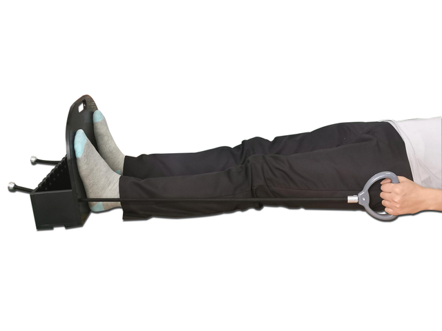
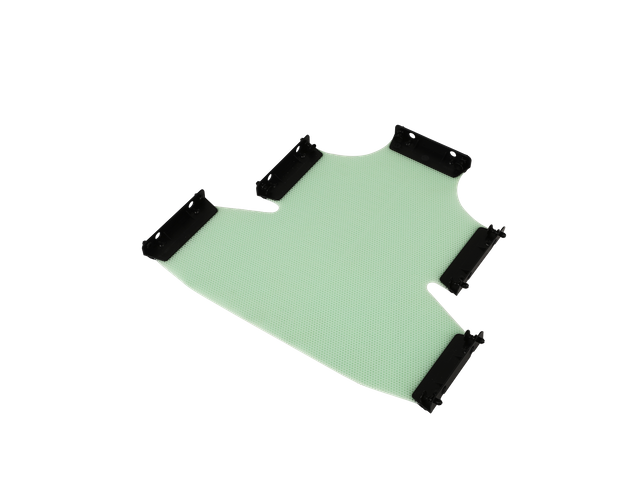
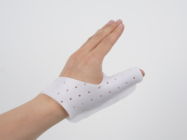
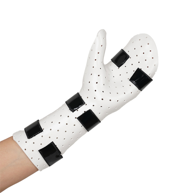
Technicians fit you with custom immobilization devices, such as thermoplastic masks, vacuum cushions, or carbon fiber breastboards. These devices prevent movement and ensure you stay in the same position for every session. Shenzhen Tengfei Yu Technology Co., Ltd. offers advanced solutions like AIO Baseplates and orthotic thermoplastics to support this step.
Your care team applies temporary or permanent marks to your skin or immobilization device. These marks guide the alignment of radiation beams and help reproduce your position each day. Sometimes, small tattoos are used for long-term accuracy.
The ct sim procedure involves scanning the treatment area with the ct simulator. You must remain still while the machine acquires detailed images. The team monitors you from outside the room and communicates through an intercom. The process does not deliver radiation therapy; it only maps your anatomy.
Once ct simulation ends, you can resume normal activities. Your care team reviews the images and uses them to design your treatment plan. They schedule follow-up appointments and explain the next steps. The marks and immobilization devices remain in place for future sessions.
Ct simulation ensures every radiation treatment matches the plan, improving safety and effectiveness.
When you experience ct simulation, you benefit from advanced technology designed for precision and safety. Modern ct simulators offer several features that set them apart from standard diagnostic ct systems. These features help your care team plan and deliver radiation therapy with high accuracy.
Large bore design: The ct simulator uses a wide opening (often 70 cm) to accommodate you and any immobilization devices, such as thermoplastic masks or vacuum cushions. This design improves comfort and allows for a variety of treatment positions.
Flat tabletop: The table matches the one used during your actual treatment, ensuring consistency between simulation and therapy sessions.
Laser alignment systems: External lasers project reference lines onto your body, helping your team align you precisely for each session.
3D volumetric imaging: The ct simulator captures detailed images in multiple planes, allowing your doctors to view your anatomy from different angles.
Multiplanar reconstruction: Your care team can examine your anatomy in coronal, sagittal, and axial views, improving tumor localization.
Digitally reconstructed radiographs (DRRs): The system generates virtual X-ray images from your ct data, which your team uses to verify treatment fields.
Adjustable slice thickness: The ct simulator can scan with thin slices (as small as 2 mm) for high detail or thicker slices for faster scans.
Dose-volume histogram analysis: Your doctors use this tool to compare different treatment plans and choose the safest option for you.
Noncoplanar beam planning: The system allows for complex beam arrangements, helping to spare healthy tissue while targeting the tumor.
4D ct imaging: Some ct simulators track tumor movement during breathing, which is important for treating tumors in the chest or abdomen.
Shenzhen Tengfei Yu Technology Co., Ltd. offers the CT Sim system, which integrates these advanced features. Their product supports a full range of immobilization devices, such as AIO Baseplates, Carbon Fiber Breastboards, and thermoplastic masks. This integration ensures you remain in the correct position throughout your treatment, improving both accuracy and comfort.
Note: Ct simulation systems focus on treatment planning, not diagnosis. The images may look different from those of a standard ct scan, and your position will match the one used during therapy.
Ct simulation plays a central role in your radiation therapy journey. The process begins when the ct simulator acquires detailed images of your treatment area. These images serve as the foundation for your personalized treatment plan.
Component | Description |
|---|---|
CT Scanner | Captures cross-sectional images for 3D planning and tumor localization. |
Image Processing Workstation | Allows your care team to outline the tumor and healthy tissues. |
Laser Marking System | Projects precise lines to guide your positioning for every session. |
Treatment Planning Software | Calculates radiation fields, beam angles, and dose distribution. |
Clinical Evaluation | Ensures accuracy and reproducibility through tests and quality checks. |
Your care team uses the ct simulation data to contour the tumor and nearby organs. They then design radiation beams that target the tumor while sparing healthy tissue. The system supports interactive 3D planning, so your doctors can adjust the plan if your anatomy changes during treatment. Advanced ct simulators also allow for adaptive planning, which means your team can update your plan if needed.
Integration with hospital information systems streamlines your experience. Your demographic and clinical data automatically transfer to the ct simulator, reducing errors and saving time. The combination of precise imaging, advanced planning tools, and reliable immobilization devices ensures you receive safe, effective, and personalized care.
Tip: Ask your care team how ct simulation and treatment planning work together to create the best possible outcome for you.
You will notice clear differences in the purpose of a ct scan compared to a ct simulation.
A ct scan serves as a general imaging procedure. Doctors use it to diagnose a wide range of medical conditions across different parts of your body.
A ct simulation is a specialized process designed for radiation therapy planning. You will encounter this if you need treatment for cancer.
During ct simulation, your care team creates immobilization devices to keep you still during therapy.
The images from ct simulation capture your body in the exact position required for radiation treatment.
Your team places targeting marks on your skin to guide the radiation beams.
The ct simulator includes advanced features like a large bore, flat tabletop, laser positioning, and virtual simulation software.
In summary, a ct scan focuses on diagnosis, while a ct simulation supports precise planning and delivery of radiation therapy.
When you prepare for a ct scan, you usually remove metal objects and change into a gown. The scan itself is quick and does not require special positioning. You lie on a table, and the machine takes images as you breathe normally. After the scan, you can return to your daily activities.
A ct simulation involves more detailed preparation. Your care team positions you exactly as you will be during radiation therapy. They may use devices like breast boards or vacuum cushions to keep you still. Sometimes, you need to hold your breath for short periods. The team may use respiratory tracking systems to match your breathing with the scan. These steps help ensure that your anatomy stays consistent for every treatment session. The team also places marks on your skin or immobilization device to guide future therapy.
A standard ct scanner produces detailed 3D images of your internal organs, bones, and blood vessels. Doctors use this equipment mainly for diagnosis.
A ct simulator includes a ct scanner but adds several specialized tools. You will find a flat tabletop, laser positioning devices, and a large bore to fit immobilization devices. The system also features virtual simulation software and a treatment planning workstation.
The ct simulator supports the creation of custom immobilization devices. These devices help you stay in the same position for each treatment session.
The equipment in ct simulation allows your care team to mark your body and plan radiation beams with high accuracy.
Tip: If you have questions about the equipment or process, ask your care team to explain how each step helps with your diagnosis or treatment.
When you prepare for a ct scan, you usually experience a quick and straightforward process. The staff asks you to remove metal items and change into a gown. You lie on a curved table, and the scan takes only a short time. The team may use a contrast agent, but you rarely need special positioning. Most people find the procedure comfortable and easy to complete.
A ct simulation feels different. You notice the table is flat and firm, just like the one used for your radiation therapy. The team spends extra time positioning you with great care. They may use special devices, such as masks or cushions, to keep you still. You might receive small marks or tattoos on your skin to help guide your treatment. The process takes longer than a standard ct scan because accuracy is critical for your therapy.
You may need to hold your breath or stay in one position for several minutes. The team checks your alignment with lasers and marks. This careful setup ensures that every treatment session matches the plan. While the simulation adds extra steps and time, it helps your doctors deliver radiation safely and precisely.
Here is a summary of how your experience may differ:
Aspect | CT Simulation Scan | Diagnostic CT Scan Planning |
|---|---|---|
Purpose | Dedicated scan for radiation therapy planning to optimize treatment accuracy and reduce toxicity | Pre-existing diagnostic scan used for planning to potentially expedite treatment |
Patient Positioning | Flat simulation CT bed, similar to treatment couch, ensuring reproducible positioning | Rounded diagnostic CT bed, may cause slight differences in tumor positioning and anatomy |
Timing | Usually scheduled separately; mean 1.9 days from simulation to first treatment, potentially delaying treatment start | Can be used if recent (ideally within 4 days), allowing treatment to start earlier by approximately 1.9 days on average |
Dosimetric Coverage (PTV) | Mean coverage ~98.8% of PTV receiving 95% prescription dose | Mean coverage ~95.4%, with slight trend toward lower coverage, especially if diagnostic CT is older or for lung lesions |
Tumor Volume Changes | Anatomy corresponds to day one of treatment, stable during planning | Tumor volume may be smaller or different due to time gap; tumor growth can affect accuracy if CT is older than 4 days |
Organ at Risk (OAR) Dose | Standard planning with constraints met | Comparable OAR doses, on average 1.4% less mean radiation dose, constraints met |
Logistical Burden | Requires additional scheduling, patient transport, and coordination, potentially increasing hospital stay and cost | Bypasses simulation step, reducing hospital stay, logistical burden, and potentially cost |
Accuracy Considerations | More reproducible positioning; less uncertainty | Requires daily image guidance (e.g., cone-beam CT) to monitor positioning and tumor changes |
Special Considerations | Standard of care for decades; tailored for treatment accuracy | Suitable especially for spine treatments or when diagnostic CT is recent; less ideal for lung lesions |
Note: CT simulation provides a more controlled and reproducible setup for your treatment. Using a recent diagnostic ct scan can speed up your care, but your team must monitor your position and tumor changes closely.
You can see the technical and clinical differences between ct simulation and diagnostic ct scans in the table below. These differences affect your comfort, the accuracy of your treatment, and the overall experience.
Aspect | CT Simulation (Radiotherapy) | Diagnostic CT Scan (Radiology) |
|---|---|---|
Bore Size | Larger bore (80–90 cm) to accommodate positioning devices | Standard bore (~70 cm) |
Tabletop | Flat tabletop for reproducible patient positioning | Curved or standard tabletop |
External Positioning Aids | External wall lasers and radio-opaque markers for alignment | Not typically included |
Scanning Field of View (SFOV) | Larger SFOV (50–70 cm) to include patient and accessories | Smaller SFOV (~50 cm) |
Reconstructed Field of View (RFOV) | Larger RFOV (60–85 cm) for treatment planning | Smaller RFOV |
Image Quality Parameters | Focus on spatial resolution, low contrast detectability, image uniformity, contrast-to-noise ratio optimized for RT | Optimized for diagnostic image quality and dose minimization |
Dose Management | Balances higher dose for better image quality in RT planning | Minimizes dose for broad patient population |
Reconstruction Techniques | Use of iterative reconstruction (IR) to optimize image quality and dose | IR also used but protocols differ |
Protocol Standardization | Challenging due to vendor differences; standardization efforts ongoing | Protocols standardized for diagnostic purposes |
Patient Positioning | Critical; requires reproducibility and inclusion of skin marks | Less critical; diagnostic positioning |
Imaging Purpose | Treatment planning, tumor and organ delineation | Diagnosis, screening, follow-up |

Tip: If you have questions about your ct experience, ask your care team to explain each step and how it helps your treatment.
You will likely encounter a ct scan when your doctor needs to diagnose or evaluate a medical condition quickly and accurately. This imaging method stands out for its ability to provide detailed views of bones, organs, and blood vessels. For example, ct scans are the preferred choice for assessing spinal injuries, such as dislocations or burst fractures, because they deliver critical information for surgical planning. Doctors also rely on ct to detect kidney stones, especially in older patients or those with unusual symptoms. In emergency situations, ct angiography helps identify ruptured abdominal aortic aneurysms, which require rapid diagnosis and treatment.
You may also need a ct scan if your doctor suspects certain cancers. This technology excels at detecting tumors in the abdomen, such as those in the stomach, esophagus, or rectum. It can also monitor changes in tumor size during treatment and spot lesions in organs like the bladder or skeleton. When bone detail is essential, such as in cases of craniosynostosis, ct provides better characterization than MRI. These strengths make ct a valuable tool for both routine and urgent diagnostic needs.
Tip: If your doctor recommends a ct scan, ask how the results will guide your care.
You will experience ct simulation if you need radiation therapy for cancer. This process creates a precise map of your body in the exact position required for treatment. The main goal is to help your care team plan and deliver radiation with high accuracy, protecting healthy tissue while targeting the tumor. During ct simulation, you will use immobilization devices, such as vacuum bags or indexed boards, to ensure you stay in the same position for every session.
Ct simulation is especially important for complex or curative radiotherapy plans. For brain tumors and thoracic lesions, reproducible positioning is critical. The images from ct simulation allow your doctors to outline the tumor and calculate the radiation dose with confidence. In some urgent palliative cases, your team may use a recent diagnostic ct scan instead of a full simulation, especially for simple plans involving bone or soft tissue metastases. However, ct simulation remains the standard for most treatment planning because it ensures accuracy and safety.
Note: Immobilization and reproducibility are key benefits of ct simulation, making it essential for many cancer treatments.
You and your care team will decide between a ct scan and ct simulation based on your specific needs. The main factor is the purpose of the imaging. If you need a diagnosis or your doctor wants to monitor a condition, a standard ct scan is the best choice. For treatment planning, especially in radiotherapy, ct simulation offers specialized features like flat tables, laser alignment, and custom immobilization devices.
Other factors include the complexity of your treatment plan and the location of your tumor. For brain tumors or areas where precise targeting is necessary, ct simulation provides the reproducibility and accuracy required. In palliative cases, where speed is important and the plan is simple, a recent diagnostic ct scan may suffice. Your care team also considers equipment availability, the need for quality assurance, and how incidental findings will be managed.
Purpose of imaging (diagnosis vs. treatment planning)
Tumor location and complexity
Need for immobilization and reproducibility
Equipment and staff expertise
Institutional policies for incidental findings
Remember: Your care team will choose the imaging method that best supports your diagnosis or treatment goals.
You can take several steps to ensure a smooth and safe ct scan experience. Your care team may ask you to fast for a few hours before the scan, especially if you need contrast dye. This helps reduce nausea and improves image clarity. Always inform your medical team about any allergies, medications, or if you are pregnant. Wear comfortable, loose-fitting clothing and remove all metal objects, such as jewelry or watches, to prevent interference with the images. If your scan requires contrast, you may need to sign consent forms and possibly undergo a blood test to check kidney function. During the scan, follow instructions to remain still and breathe as directed. This helps produce clear images for your doctor.
Tip: Ask questions if you feel unsure about any part of the process. Understanding each step can help reduce anxiety.
Preparing for ct simulation involves a few unique steps compared to a standard scan. Your care team will review your medical history, previous imaging, and treatment plans before the appointment. You may receive specific instructions, such as emptying your bladder or keeping it full, depending on the treatment area. Psychological preparation is important, so your team will explain the need for staying still and the use of skin marks or small tattoos. Specialized immobilization devices, like thermoplastic masks or vacuum cushions, help keep you in the same position for every treatment session. The team will mark your skin or the device with reference points to guide radiation targeting. They also verify that all necessary equipment and documentation are ready before starting.
Review your treatment plan and ask about any special instructions.
Wear comfortable clothing and remove metal items.
Prepare for possible skin marking or tattooing.
Understand the importance of remaining still during the simulation.
Note: The simulation process may take longer than a standard scan, but it ensures your treatment is as accurate as possible.
After your ct scan or simulation, you can usually return to normal activities. If you received contrast dye, drink plenty of fluids to help flush it from your system. Watch for any signs of allergic reaction, such as rash or difficulty breathing, and contact your care team if you notice anything unusual. For ct simulation, your team will schedule your set-up procedure and treatment sessions. Radiation treatments often occur Monday through Friday, and attending every session is important for the best results. If you cannot make an appointment, notify your care team as soon as possible. Before your first treatment, the team will position you exactly as during simulation and take special x-rays to confirm accuracy. You can take vitamins and multivitamins during therapy, but avoid exceeding recommended daily amounts. Discuss any supplements with your care team.
Your radiation oncologist and nurse remain available to answer questions and support you throughout your treatment and recovery.
When you prepare for a CT scan or CT simulation, you should feel empowered to ask your care team important questions. Open communication helps you understand each step and reduces anxiety. Here are some key questions you might consider:
What specific preparation do I need before my scan or simulation? Should I follow any diet restrictions or take certain medications?
Will I need to empty or fill my bladder or bowels before the procedure?
Can you explain the purpose of any immobilization devices or tattoos used during the simulation?
What should I expect during the procedure? Will I need to wear a gown or remove jewelry?
How will my appointments be scheduled? When will my simulation and treatment sessions take place?
Are there any side effects I should watch for? What support services are available if I need help?
Do I need a pregnancy test before the simulation?
How can I manage transportation and support during my treatment?
What skin care instructions should I follow? How do I handle any skin reactions?
Tip: Write down your questions before your appointment. Bringing a list helps you remember what to ask and ensures you get the information you need.
After your scan or simulation, you will receive results that guide your care. Understanding these results helps you stay involved in your treatment plan. CT simulation scans use special equipment and positioning to match your treatment setup. This approach allows your care team to target tumors accurately and protect healthy tissue. Diagnostic CT scans, on the other hand, do not use these aids. They may not reflect your treatment position, which can affect the precision of your radiotherapy plan.
You can compare different planning methods in the table below:
Method | Advantages | Disadvantages |
|---|---|---|
Simulator-CT | Best for covering the target and sparing normal tissues | Takes more time |
Surface marking | Simple and quick | May not cover the target well; higher dose to healthy organs |
Simulator fluoroscopy | Middle ground between surface marking and Simulator-CT | Acceptable in some cases |
Planning CT scans often show smaller target areas than diagnostic scans. This difference happens because your body position changes between scans. Target areas can shift, which affects accuracy. Adaptive treatment plans, based on images taken during your therapy, help your team cover the tumor better and protect healthy organs. If your plan uses only a diagnostic CT, your team may need to adjust it to make sure the radiation covers the tumor completely.
Note: Always ask your care team to explain your results in simple terms. If you do not understand something, ask for clarification. Your involvement helps ensure the best possible outcome.
You now know that a CT scan helps diagnose medical conditions, while a CT simulation prepares you for precise radiation therapy.
Use a CT scan when your doctor needs detailed images for diagnosis.
Choose CT simulation when you need accurate treatment planning for cancer care.
Stay informed and ask your care team questions. Your understanding and involvement help ensure the best possible outcome for your health.
You use a CT scan for diagnosis. You use a CT simulation for planning radiation therapy. CT simulation positions you for treatment and helps your care team target tumors accurately.
You may need to follow instructions about eating, drinking, or emptying your bladder. Your care team will explain how to prepare and what to expect during the simulation.
You do not receive treatment radiation during CT simulation. The scan only creates images for planning. You start actual radiation therapy after your care team completes your plan.
Immobilization devices keep you in the same position for every treatment. These devices help your care team deliver radiation precisely and protect healthy tissue.
Your care team may use a recent diagnostic CT scan for simple cases. For most treatments, CT simulation provides better accuracy and reproducibility.
CT simulation usually takes 30 to 60 minutes. You spend extra time on positioning and marking to ensure your treatment matches the plan.
You may receive temporary marks or small tattoos. These marks help your care team align you for each radiation session.
Your radiation oncologist, nurse, or technologist can answer your questions. You should ask about preparation, procedure, and aftercare.
 English
 简体中文
简体中文